
Tilapia skin in dogs has been studied in protocols related to canine ophthalmology and ocular regeneration.
This content is intended exclusively for informational purposes and does not replace professional veterinary evaluation.
Veterinary medicine has advanced significantly in the development of biomaterials aimed at tissue regeneration and healing support across different clinical fields. Among the topics that have attracted growing scientific interest in recent years is the use of tilapia skin in dogs within research related to veterinary ophthalmology.
Initially studied in contexts associated with tissue regeneration, tilapia skin drew researchers’ attention due to its high collagen content and biological characteristics considered favorable for regenerative applications. Based on these observations, studies began investigating possible applications of this biomaterial in canine ophthalmology, especially in protocols related to corneal recovery and ocular surface protection.
Why Has Tilapia Skin in Dogs Begun to Attract Scientific Interest?
Tilapia skin presents structural characteristics that have been carefully observed in medical and veterinary research. Among the aspects most frequently discussed in studies are:
- high natural collagen concentration;
- biologically resistant structure;
- moisture-retention capacity;
- temporary tissue protection;
- regenerative support potential;
- biological compatibility in specific applications.
These properties led Brazilian researchers to investigate tilapia skin in dogs in different regenerative contexts, including ophthalmological applications involving the cornea.
What Is the Cornea and Why Do Alterations in This Region Require Attention?
The cornea is the transparent structure located at the front of the eyes and plays an essential role in ocular protection and proper light transmission. When alterations occur in this region, discomfort can become significant and require specialized veterinary monitoring.
Among the signs that frequently draw tutors’ attention are:
- ocular redness;
- frequent itching;
- excessive tearing;
- light sensitivity;
- presence of discharge;
- difficulty keeping the eyes open;
- persistent discomfort.
Some breeds, especially brachycephalic dogs, tend to present a greater predisposition to ophthalmological alterations due to their more exposed ocular anatomy. These include Shih-tzu, Lhasa Apso, Bulldog, and Pug.
How Has Tilapia Skin in Dogs Been Studied in Veterinary Ophthalmology?
In canine ophthalmology, tilapia skin has been analyzed as a possible auxiliary biomaterial in specialized protocols aimed at ocular surface recovery. In certain experimental and clinical approaches, the material may function as a temporary biological covering applied over injured areas of the cornea.
The purpose of these applications is not to replace conventional veterinary treatments, but rather to assist local protection and provide favorable conditions for regenerative processes involving the ocular surface.
Among the studied applications of tilapia skin in dogs are:
- regenerative support for the cornea;
- temporary protection of the ocular surface;
- support for healing processes;
- maintenance of local moisture;
- tissue protection in specific ophthalmological alterations.
Brazilian Research and Veterinary Regenerative Medicine
Interest in tilapia skin in dogs gained even greater visibility because it involves research developed in Brazil. The topic attracted attention within regenerative medicine due to the biological potential of the material and its possible applications across different clinical fields.
In veterinary ophthalmology, cornea-related studies have drawn interest precisely because they seek alternatives capable of supporting tissue recovery in certain ophthalmological situations.
Although many protocols are still associated with specialized contexts and ongoing research, the observed progress demonstrates the growth of regenerative approaches within contemporary veterinary medicine.
In Our Family Context
Within our family context, we recently experienced ocular irritation involving a 12-year-old Shih-tzu named Luna, a situation that further increased our interest in advances in veterinary ophthalmology and research related to tilapia skin in dogs.
From this experience onward, we began following scientific studies and content related to ocular recovery and new regenerative possibilities focused on veterinary ophthalmological care.
What Advantages Have Researchers Observed in This Biomaterial?
Among the factors most frequently observed in research involving tilapia skin in dogs are:
- high collagen presence;
- adaptation to the ocular surface;
- temporary biological protection;
- maintenance of tissue moisture;
- regenerative potential;
- resistant and flexible structure.
In addition, the subject has received attention precisely because it combines regenerative medicine, biomaterials, and advances in veterinary ophthalmology.
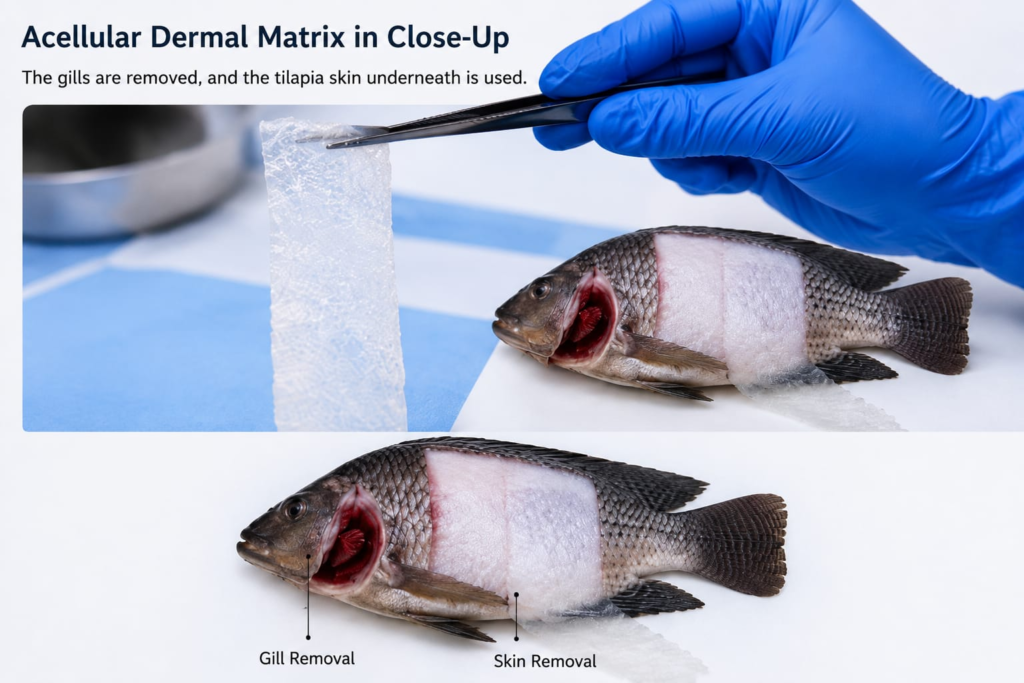
Preparation process of tilapia skin used in veterinary regenerative research.
Where Is Tilapia Skin Used in Veterinary Studies Prepared?
It is important to understand that the tilapia skin used in medical and veterinary studies is not the same skin intended for ordinary food consumption. The material employed in specialized protocols undergoes specific processes, including:
- sterilization;
- laboratory processing;
- microbiological control;
- rigorous biosafety protocols;
- specialized technical preparation.
For this reason, any application related to tilapia skin in dogs depends exclusively on veterinary evaluation and specialized professional supervision.
Applications in More Complex Ophthalmological Situations
In certain more delicate ophthalmological contexts, researchers have also been observing possible uses of tilapia skin in dogs involving deeper corneal alterations. In these situations, factors such as lesion extent, inflammatory response, tissue involvement, and time of progression become fundamental for any therapeutic decision.
Each ocular condition requires individualized evaluation, especially in dogs presenting significant visual alterations or more advanced ocular surface impairment.
To further explore this topic, also read this related post: https://logicalbark.com/is-my-senior-dog-going-blind-signs-causes-and-essential-care/
The Advancement of Veterinary Ophthalmology
The growing interest in tilapia skin in dogs demonstrates how veterinary medicine has been expanding research focused on tissue regeneration and the development of biomaterials applied to canine ophthalmology.
Although many studies are still under development, the observed advances reveal a promising scenario for future regenerative approaches within specialized veterinary medicine.
In the presence of persistent signs such as redness, ocular irritation, discharge, or continuous discomfort, early veterinary evaluation remains essential to preserve ocular health and animal well-being.
In certain ophthalmological contexts, tutors also seek products aimed at supporting ocular hydration, cleaning, and visual comfort in dogs. Among the options available in the veterinary market are lubricating eye drops and specific solutions developed for animal use.
Some examples include products intended for ocular lubrication and veterinary eye support, always used exclusively under proper professional guidance: https://amzn.to/4cUbdxQ
Disclaimer: This content is intended exclusively for informational and educational purposes. Veterinary ophthalmological procedures, biomaterials, and ocular treatments must be evaluated and conducted only by specialized veterinarians. Never perform ocular interventions without proper professional guidance.
You may also like

Lusiane Costa is a digital writer with degrees in Marketing and English Literature.
Creator of Latido Lógico and Logical Bark, she develops evidence-based content on canine aging, wellness, and senior-dog health.
The project was inspired by Goe — a senior dog whose longevity and resilience shaped a grounded, compassionate view on the challenges of aging in pets.
Each article reflects her commitment to transforming real experiences into accessible knowledge, helping owners understand, prevent, and care better for their animals at every stage of life.
Goe remains the heartbeat of this project.